Epilepsy
As a graduate student, I worked with the labs of Mark Kramer and Uri Eden at Boston University and Sydney Cash at Massachusetts General Hospital to analyze human seizures. In particular, we focused on analyzing how seizures stop with microelectrode array (MEA) data, below.
Seizure A1
Statistical Models
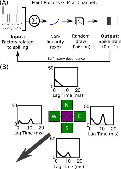
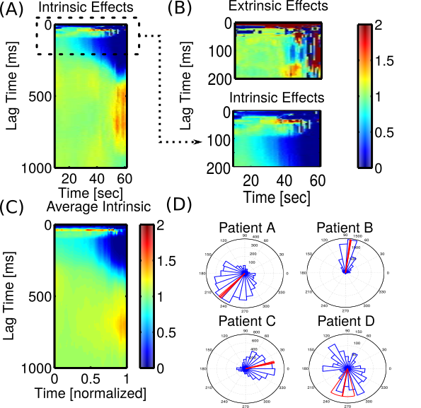
We use statistical models to describe how functional networks interact to produce "ictal discharges" (IDs): pathological events in the extracellular electric field field during seizure. IDs exhibit rich spatiotemporal structure, as shown in the movie above.
Importantly, IDs are point process data: discrete counts isolated in time and space. A rigorous and powerful approach to analyzing such data is the point process generalized linear model (PP GLM). The PP GLM below parses within-channel ("intrinsic") and cross-channel ("extrinsic") dynamics simultaneously. We find the typical rhythmic bursting associated with the end of spike-wave seizures, as well as strong evidence that IDs propagate with a preferred direction with respect to the seizure onset zone.
Biophysical Models
I have also worked on simulating more detailed biophysical models, such as the leaky integrate-and-fire neuron. Pictured below is work done with Andrew Sornborger to complement calcium imaging of zebrafish undergoing chemically-induced seizures. The video shows a network of 30x100 LIF neurons stimulated with an applied current at the left edge of the grid. Because the range of inhibitory connections is less than normal (top), the stimulation produces a spatial wave of spiking. With longer inhibitory connections (bottom), such a wave fails to propagate. The model is based on work by Ursino & La Cara (2005), and similar work has been done by Hall & Kuhlmann (2013).
Excitation produces a spatial wave of spiking
Feedforward inhbition restrains a spatial wave